The transmission electron micrographs included in this collection were all the work of Dr. Eldon Newcomb. Dr. Newcomb was a pioneer in the fixing and preparation of plant tissue for viewing with the transmission electron microscope. Dr. Newcomb generously allowed me to scan a sampling of his glass plates on a flat bed scanner. I do not have documentation about the source of the tissue scanned. However, each of these examples illustrates good generic views of plant cellular ultrastructure.
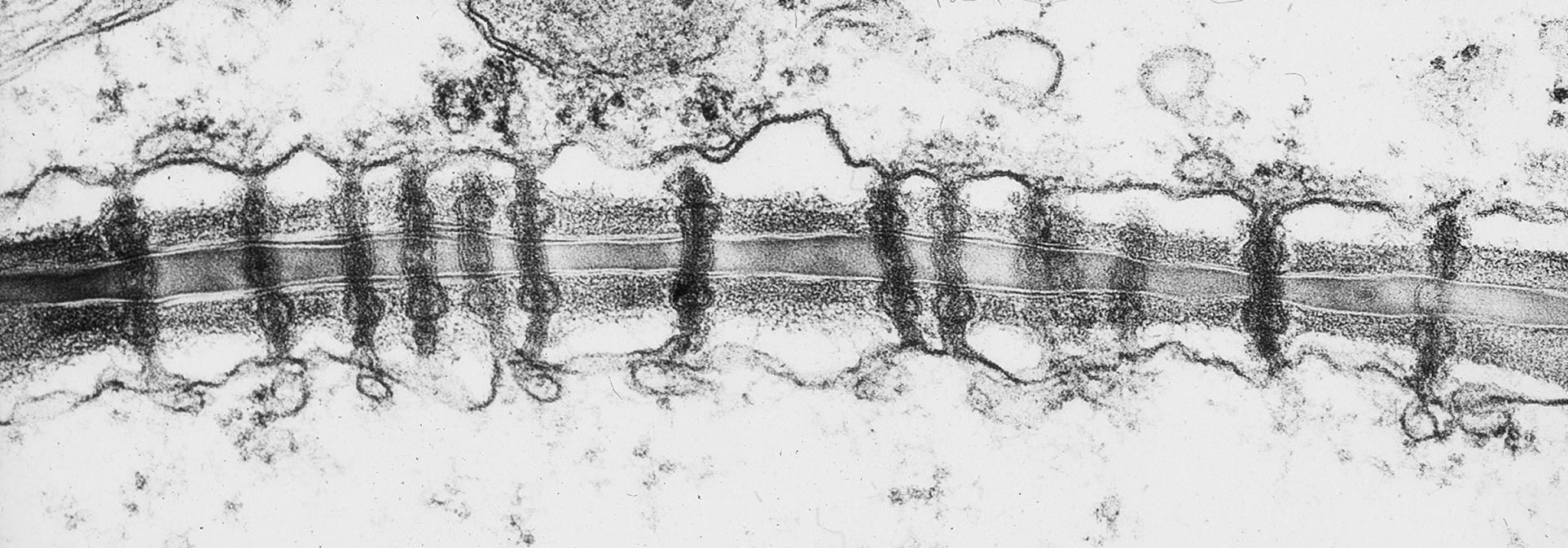
This section was made parallel to the cell wall and cut through it and through part of the underlaying cytoplasm. This section shows plasmodesmata in longitudinal section. These channels of cytoplasm connect adjacent plant cells together. They are barely visible in longitudinal section with a light microscope (see light microscope images of Diospyros endosperm).