The transmission electron micrographs included in this collection were all the work of Dr. Eldon Newcomb. Dr. Newcomb was a pioneer in the fixing and preparation of plant tissue for viewing with the transmission electron microscope. Dr. Newcomb generously allowed me to scan a sampling of his glass plates on a flat bed scanner. I do not have documentation about the source of the tissue scanned. However, each of these examples illustrates good generic views of plant cellular ultrastructure.
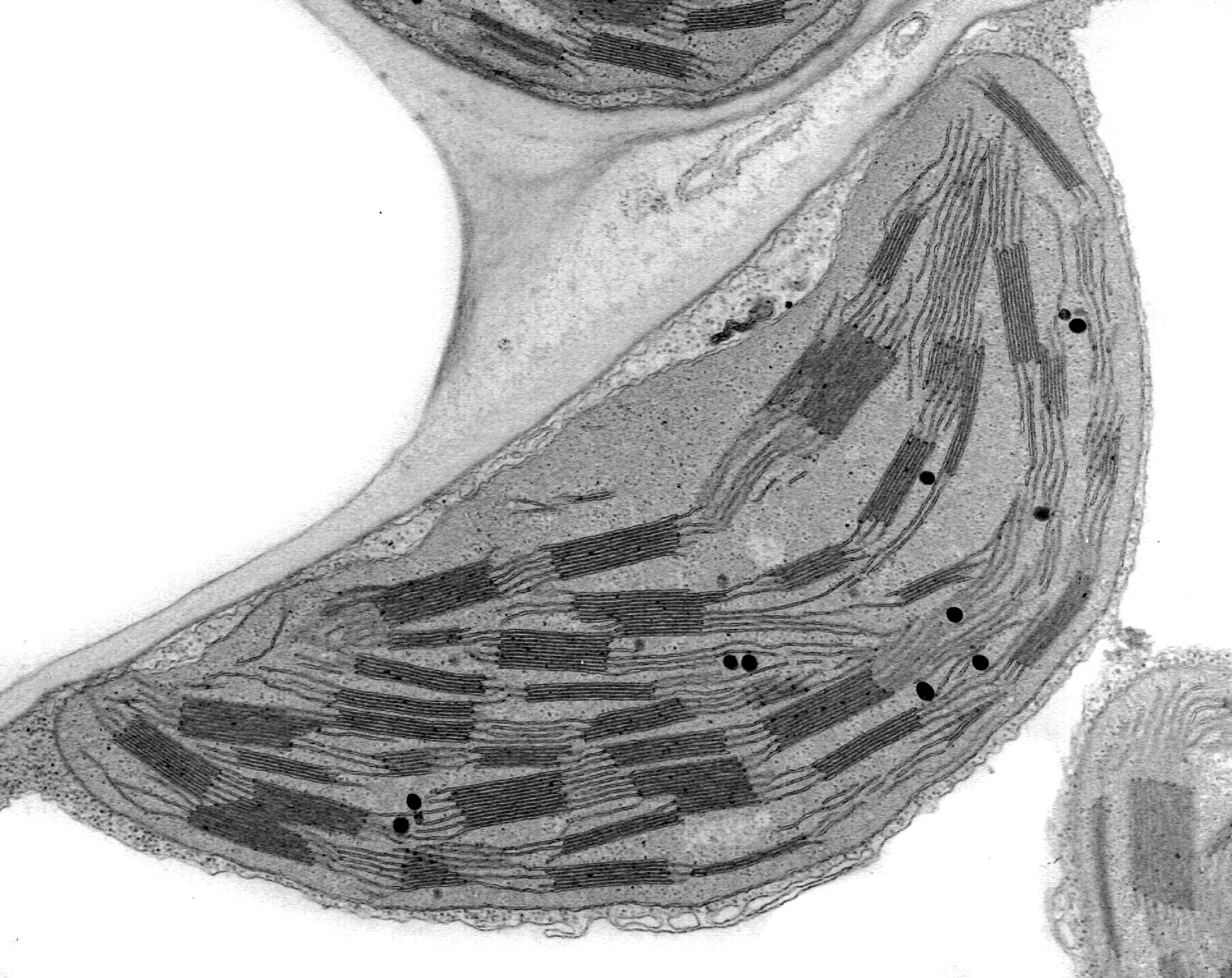
This is a detailed view of the internal structure of a chloroplast. Chloroplasts have membranes called thallakoids embedded in a ground area termed stroma. These membranes contain the two photosystems. This structure is similar to the internal structure of cyanobacteria. In chloroplasts, and unlike cyanobacteria, these tallakoids form layered structures called grana.
